 When a person goes to an eye doctor appointment, they may receive a prescription after their exam. This prescription is made up of an odd series of letters and numbers that are instructions for making your glasses. Here is what they mean:
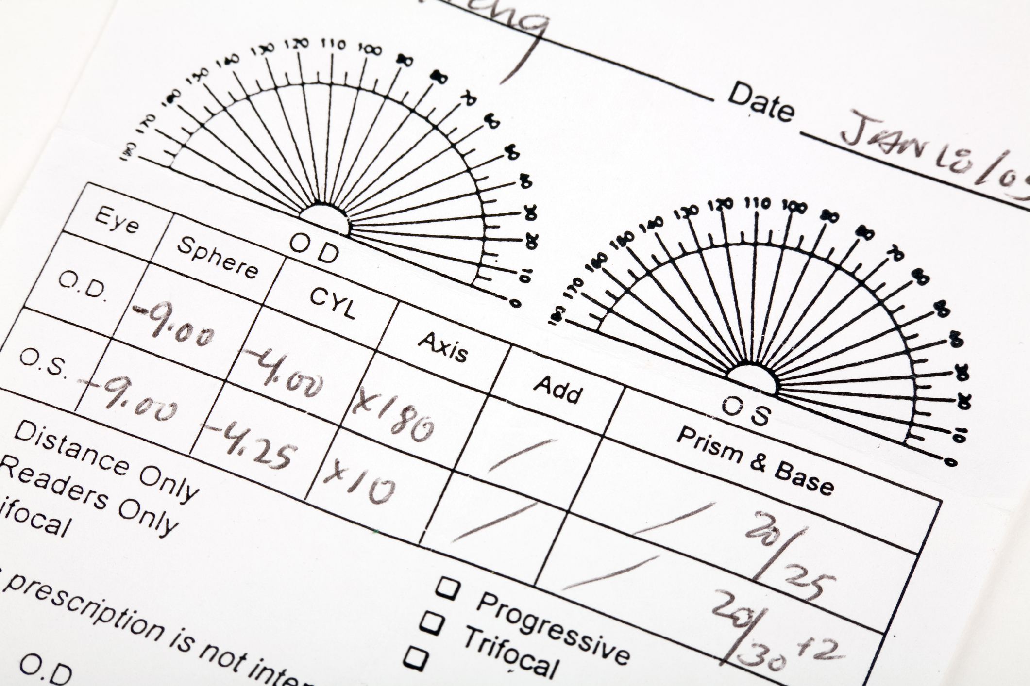
In a typical prescription, there are two acronyms, one for each eye.
- D.- is short for oculus dexter, which is your right eye
- S.- is short for oculus sinister, which is your left eye
The sphere column is often abbreviated as SPH. This is the lens power needed to fix your vision. A minus sign (-) next to the number means nearsightedness. This means you see better up close and need distance correction. A plus sign (+) indicates that you are farsighted and can see better from far away and need your near vision corrected.
Lens power is measured in diopters, the unit of measurement used to calculate the focusing strength of a pair of glasses or contact lenses. If you see the sphere field written above as -9.00 D, this means there are 9 diopters of nearsightedness. The measuring system is an integer line, with zero in the middle, needing no correction. The further you get away from zero on either the minus or plus side, the stronger your prescription is.
The cylinder number is how much astigmatism you have, if any. This is when part of the cornea has a different curve. Normally, an eye is shaped like a basketball, rotated in any direction, with the curve staying the same. An eye with astigmatism is oval, or egg-shaped, or more like a football, with one curve being longer than the other. The CYL number corrects the different second curve.
The axis number tells you where the astigmatism is on the cornea. The axis is written in degrees between 1 and 180, indicating which way the astigmatism lines up.
The add column is where any additional lens power is written. For example, some people over the age of 40 may not want an extra pair of glasses for reading and may choose to wear bifocals instead. The lower half of the lens will give them their reading vision.
Additionally, there may be a field for prism on the right side. This is a special type of correction built into the lens for some people with double vision. This means they see two separate images of the same object. The prism fuses the two images together so they will only see one image.
The prescription for contact lenses is different because they sit directly on the eye. A contact lens prescription includes measurements specific to the size and brand of your contacts. Before you fill a prescription for contact lenses, you will need a contact lens fitting to see if they are right for you.
If you require an eye exam, you can schedule an appointment at Jamaica Hospital Medical Center’s Ophthalmology Center by calling (718) 206-5900.
 Follow
All content of this newsletter is intended for general information purposes only and is not intended or implied to be a substitute for professional medical advice, diagnosis or treatment. Please consult a medical professional before adopting any of the suggestions on this page. You must never disregard professional medical advice or delay seeking medical treatment based upon any content of this newsletter. PROMPTLY CONSULT YOUR PHYSICIAN OR CALL 911 IF YOU BELIEVE YOU HAVE A MEDICAL EMERGENCY.


 A s
A s